Dr. Stefan Scherg

Peri-implantitis treatment with GalvoSurge®

Single implant, closed healing

Dr. Stefan Scherg

Germany


Case courtesy of Dr. Scherg

Patient: 42-year-old male, nonsmoker, no history of periodontitis, implant placed 3 years ago.

Clinical situation: Single implant in FDI tooth position 36. Crater-like bone defect, RP classification 1(-2), with 6 mm bone loss due to advanced peri-implantitis. Visual signs of inflammation, PPD of 8 mm, BoP. The patient wants to keep the implant because it is relatively new.

Treatment plan: Removal of the prosthesis, presurgical periodontal and anti-infective therapy and placement of a cover screw 2 weeks before the surgical intervention. Due to the position of the implant close to the mandibular nerve, block anesthesia was sed (Articain, 1.8 ml). In surgery, the opening of a full-thickness flap, removal of the granulation tissue with curettes, no hard deposits to be removed, and removal of the biofilm using the GalvoSurge Dental Implant Cleaning System. Placement of a cover screw. Guided bone regeneration using bovine bone mineral (creos™ xenogain) and collagen membrane (creos™ xenoprotect). Medication with antibiotics (Augmentan 875/125mg 1-0-1). Closed healing for 3 months. Placement of a healing abutment 3 months after surgery, and placement of the existing prosthesis (cleaned and sterilized) 4 months after surgery.

Patient follow-up: After surgery, the patient was medicated with Amoxicillin with Clavulanic acid for 7 days and ibuprofen 600 mg. After 12 days the sutures were removed. 3 months after the surgery a healing abutment was placed, 4 months after surgery the final prosthesis was placed. Patient follow-up every 6 months. 

Surgery date: March 9, 2019

Total treatment time: 6 months


Case courtesy of Dr. Stefan Scherg.

Some products may not be regulatory cleared/released for sale in all markets.

Please use landscape mode when viewing this page on mobile.

Initial clinical situation

Case courtesy of Dr. Scherg

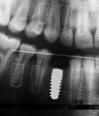
Progression of the bone loss.

Implant placement. Transmucosal healing because of implantation without bone graft (first image from left).

3-months follow-up. Initial bone loss (second image from left).

22-months follow-up. Progressive bone loss (third image from left).

Clinical picture at diagnosis. Visual signs of inflammation and suppuration (fourth image from left)

Presurgical therapy

Case courtesy of Dr. Scherg

Clinical situation after removal of the prosthesis. Visual signs of inflammation and suppuration (left).

Presurgical periodontal and anti-infective therapy and placement of a cover screw (center).

Removed prosthesis, to be cleaned, sterilized and placed back in the patient mouth after 6 months (right).

Surgical procedure

Case courtesy of Dr. Scherg

Removal of the granulation tissue and hard deposits.

Clinical situation 2 weeks after removal of the prosthesis (left).

Removal of the granulation tissue using curettes. Implant after removal of the granulation tissue (center).

Implant after removal of the granulation tissue (right).

Surgical procedure

Case courtesy of Dr. Scherg

Implant surface decontamination using GalvoSurge.

Placement GalvoSurge Spray Head on the implant. Cleaning time 2 minutes (first image from left).

Hydrogen bubbles lift the oral biofilm off the implant surface creating a clean implant surface. (second image from left).

Implant surface after cleaning with GalvoSurge (third image from left).

Placement of a sterile cover screw on cleaned implant (fourth image from left).

Surgical procedure

Case courtesy of Dr. Scherg

Guided bone regeneration using creos™ xenogain and creos™ xenoprotect.

Guided bone regeneration using bovine bone mineral (creos xenogain) (left).

Placement of a collagen membrane (creos xenoprotect) (center).

Suture. Closed healing for 3 months (right).

Healing process and outcome

Case courtesy of Dr. Scherg

4 months healing phase and final outcome.

3 months after surgery. Placement of a healing abutment (left).

4 months after surgery. Placement of the cleaned and sterilized original prosthesis (center).

12 months after surgery. Peri-implant mucosa is healthy, no BoP, PPD >2mm, no more suppuration (right).

Healing process and outcome

Case courtesy of Dr. Scherg

X-ray after surgical intervention (left).

4 months after surgery. Placement of the prosthesis (center).

12 months after surgery. Complete bone fill (right).

Related products

Sign up for our blog update

Get the latest clinical cases, industry news, product information and more.

This clinical case was prepared/accomplished in the clinician's name, in his/her personal capacity. The views, clinical opinions and scientific interpretations expressed in this video are the author's own and do not necessarily represent the ones of Nobel Biocare.
© Nobel Biocare Services AG, 2022. All rights reserved. Nobel Biocare, the Nobel Biocare logotype and all other trademarks are, if nothing else is stated or is evident from the context in a certain case, trademarks of Nobel Biocare. Please refer to nobelbiocare.com/trademarks for more information. Product images are not necessarily to scale. Disclaimer: Some products may not be regulatory cleared/released for sale in all markets. Please contact the local Nobel Biocare sales office for current product assortment and availability. For prescription use only. Caution: Federal (United States) law restricts this device to sale by or on the order of a licensed clinician, medical professional or physician. See Instructions For Use for full prescribing information, including indications, contraindications, warnings and precautions. Nobel Biocare does not take any liability for any injury or damage to any person or property arising from the use of this clinical case. This clinical case is not intended to recommend any measures, techniques, procedures or products, or give advice, and is not a substitute for medical training or your own clinical judgement as a healthcare professional. Viewers should never disregard professional medical advice or delay seeking medical treatment because of something they have seen in this clinical case. Full procedure is not shown. Certain sequences have been cut.