Ireland


CLINICAL CASE
Nobel Biocare N1™ concept in the posterior maxilla with sinus floor elevation
Dr. Oded Bahat, Dr. Ion Zabalegui,
Dr. Eva Berroeta, Mr. Javier Pérez
CLINICAL CASE
Dr. Oded Bahat, Dr. Ion Zabalegui,
Dr. Eva Berroeta, Mr. Javier Pérez

Partial edentulism right side maxilla with vertical ridge deficiency.
Healed site before surgery. Lateral view (left).
Healed site before surgery. Occlusal view (right).
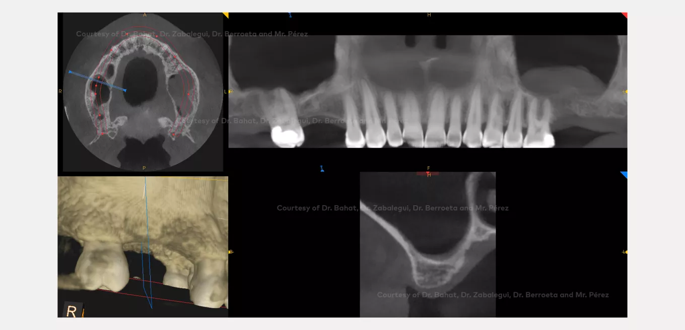
CBCT Pre-op. CBCT illustrating inadequate vertical bone height, right side and healthy sinus. Mucosal thickening left sinus.

Flap delineation and full thickness elevation.
Marking of flap (left).
Buccal view of ridge (right).
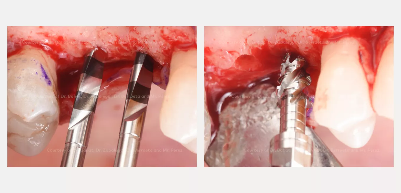
OsseoDirector™ instruments (2000 rpm with irrigation). Initial osteotomy with the OsseoDirector instrument. The final length and implant position established (left).
Osseoshaper™ instrument* preparation of the site at 50rpm without irrigation (left).
* In this clinical case the pre-launch version of the OsseoShaper™ instrument has been used: the “CS Soft Bone Gizmo”. Black markings on the shaft and a 0.5 mm longer tip on the instrument are introduced on the final design of the OsseoShaper™ instrument.

Implant followed the direction created by the OsseoDirector™ and OsseoShaper™ instruments* (left).
Flat side of the trioval implant facing buccal (right).
* In this clinical case the pre-launch version of the OsseoShaper™ instrument has been used: the “CS Soft Bone Gizmo”. Black markings on the shaft and a 0.5 mm longer tip on the instrument are introduced on the final design of the OsseoShaper™ instrument.

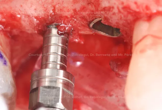
OsseoShaper™ instrument* at full depth. The end of the threads indicates the position of the implant shoulder relative to the bone crest (left).
Reversing to remove the instrument (right).
* In this clinical case the pre-launch version of the OsseoShaper™ instrument has been used: the “CS Soft Bone Gizmo”. Black markings on the shaft and a 0.5 mm longer tip on the instrument are introduced on the final design of the OsseoShaper™ instrument.

Autogenous bone is maintained within the osteotomy and within the threads of the instrument (left)
Sinus lift with osteotomes after site preparation (right).

Implant insertion 70 Ncm. Final placement of the implants. Flat part on the implant driver and vertical corresponding groove within the mount corresponds to the trioval flat aspect facing the facial aspect

Transmucosal base placed at time of surgery (left).
Healing caps and the base function as healing abutments (right).

Flap closure. Healing caps and the base jointly shape mucosa (left).
Radiographs immediately post-op (right).

CBCT immediately post-op illustrating the implants position and sinus floor.

CBCT pre-op (position 16) illustrating inadequate vertical bone height.

CBCT Post-op (position 16) illustrating implant placement and sinus elevation.

12 month post-op final restoration.

35 months post-op. Follow up (left).
CBCT 35 months post-op (right).

35 months post-op facial view.
© Nobel Biocare Services AG, 2019. All rights reserved. Nobel Biocare, the Nobel Biocare logotype and all other trademarks are, if nothing else is stated or is evident from the context in a certain case, trademarks of Nobel Biocare. Please refer to nobelbiocare.com/trademarks for more information. Product images are not necessarily to scale. Disclaimer: Some products may not be regulatory cleared/released for sale in all markets. Please contact the local Nobel Biocare sales office for current product assortment and availability. For prescription use only. Caution: Federal (United States) law restricts this device to sale by or on the order of a licensed clinician, medical professional or physician. See Instructions For Use for full prescribing information, including indications, contraindications, warnings and precautions. Nobel Biocare does not take any liability for any injury or damage to any person or property arising from the use of this clinical case. This clinical case is not intended to recommend any measures, techniques, procedures or products, or give advice, and is not a substitute for medical training or your own clinical judgement as a healthcare professional. Viewers should never disregard professional medical advice or delay seeking medical treatment because of something they have seen in this clinical case. Full procedure is not shown. Certain sequences have been cut.