
Heat and speed of dental implant drilling: How could site viability be improved?
Over the past few decades since the inception of implantology, implant system design and treatment concepts have come forward leaps and bounds. A vast choice of dental implants has evolved; however it may be surprising that some aspects have seen relatively little change. Based on biological research with a basic science approach, one such area is the effect of dental implant drilling protocols and implant site preparation. In this article, we unpack some of the biological response to conventional bone drilling and how Nobel Biocare is addressing the associated draw backs by introducing the OsseoShaper™ protocol.*
How do conventional, high-speed dental drills impact implant site viability?
Even with the most careful site preparation, it should be no surprise that inevitably, drilling into bone damages the bone tissue and the osteocytes – the cells that enable new bone formation and osseointegration – surrounding the osteotomy.1
A significant factor is the high speed that a conventional dental drill needs to operate effectively.2 The problem with high speed and friction is that they generate heat.4 This heat radiates from the cutting drill edge into surrounding tissue and kills bone cells, and bone cell death triggers bone resorption at the implant site.1,5,6,7
Ultimately, therefore, the heat generated by high-speed drilling can impede the much-coveted implant stability.5
This area of dead and dying osteocytes around the osteotomy has been termed the ‘Zone of Death’, and in triggering bone resorption, presents one of the challenges in achieving optimal site viability. What options might clinicians consider for minimizing causes of cell death?

Images courtesy of Prof. Jill Helms A thermograph of the osteotomy (left) demonstrates the heat radiation that is caused by drilling the bone. In the histologic image of the osteotomy (center) you cannot see any difference between bone at the periphery of the osteotomy and bone that is further away – however the TUNEL imaging (right) clearly shows a ring of apoptotic osteocytes (green) surrounding the osteotomy. This area of dead and dying cells that we can see in the TUNEL image is known as the Zone of Death.
Could more irrigation help a viable implant site?
To counteract the effects of heat, saline irrigation is typically applied in the osteotomy at each step of the drill sequence, cooling the temperature. Should we, then, simply increase irrigation to reduce heat further?
Unfortunately, irrigation brings its own drawbacks. When drilling to create an osteotomy, autologous bone chips and osseous coagulum – a blend of blood, bone cells and growth factors – are created. Because of their osteogenic properties, when left in situ this valuable material can promote new bone formation.8
However, copious irrigation washes away bone chips and osseous coagulum. Once the bone chips are removed from the osteotomy, cells quickly begin to die, and so, even if retrieved, their osteogenic potential quickly deteriorates.9 To maximize the healing potential of bone chips, the cell death should be minimized while maintaining bone chips in situ.
Why not just operate the drill at a lower speed?
A potential solution may have crossed your mind. If speed generates heat, and heat kills cells, why not simply operate the handpiece at a low rpm? The design of most implant drills requires high speed rotation in order to minimize the thrust force needed to advance through the bone.10 Without following the proper usage of a drill, safety and efficacy are compromised.
A site preparation technique to preserve vital bone – the OsseoShaper™ concept
It is clear, that the foundation of implantology – osseointegration – can be improved by optimizing osteotomy site preparation.2 For site viability then, based on some of the biological principles, this begs the questions:
- Can we minimize the Zone of Death and increase the number of viable cells?
- Can we reduce heat to eliminate the need for irrigation?
- Can we eliminate irrigation to retain bone chips?
Finding such solutions has inspired the Nobel Biocare OsseoShaper™ concept, a simplified and biologically driven site preparation protocol designed to preserve vital bone and thereby promote fast osseointegration.
-
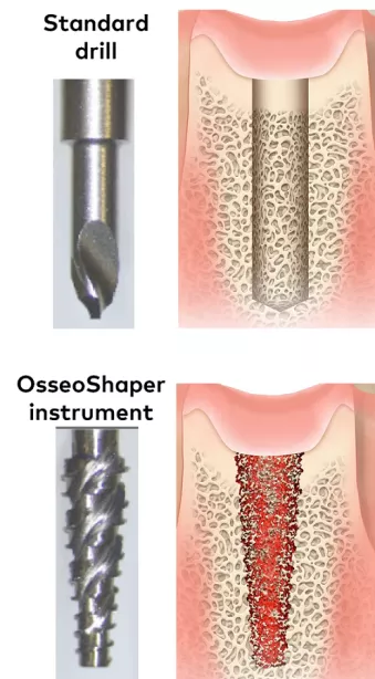
The standard drill protocol creates a smooth osteotomy (top) devoid of osseous coagulum, whereas the OsseoShaper concept (bottom) creates a textured osteotomy
-
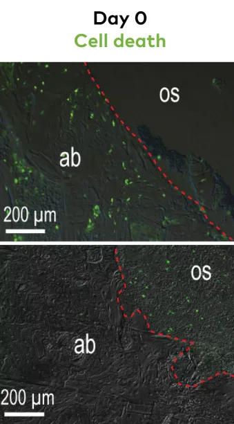
The green represents dying cells, (apoptic osteocytes). With the standard drill, a zone of dying cells are clearly visible around the osteotomy. With the OsseoShaper instrument, the majority of apoptic osteocytes are seen inside osseous coagulum, retained in the osteotomy.(9).
-
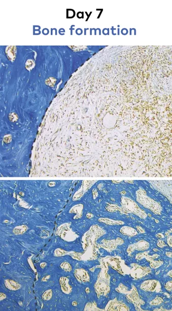
Tissue sections stained with aniline blue show minimal new bone formation in the standard drill group (above), while osteotomies in the OsseoShaper group show more new bone formation 7 days after osteotomy creation in a rodent model(9).


Developed as part of the full Nobel Biocare N1™ implant system*, the OsseoShaper instrument ‘shapes’ the osteotomy:
- It operates at low speed – just 50 rpm.
- Less heat is generated compared to high-speed drills.9
- No irrigation is required.
By eliminating irrigation, osteogenic material is preserved.9 The autologous bone chips and osseous coagulum are maintained in the osteotomy, enabling osteoinduction to occur within the osteotomy itself.
This is also assisted by the fact that many bone chips produced during site preparation are deposited into the osteotomy when the instrument is reversed. This concept has been shown to create less trauma, leading to earlier bone formation within the osteotomy compared to conventional high-speed techniques.9,11

But not only does the OsseoShaper protocol address biological principles, it has other practical benefits. Firstly, the minimally invasive site preparation is considerate of patient comfort with minimized noise and vibration. With few surgical instruments needed the concept also reduces treatment time – in fact, over 80% of recorded cases have required just two site preparation steps.12 To reduce the guesswork compared to a conventional high-speed drill protocol, and assist the clinician’s decision making during site preparation, torques generated using the OsseoShaper concept guide the surgical workflow, assist in bone evaluation and provide an early prediction of implant stability.13
References
1. Chen CH et al. A Comparative Assessment of Implant Site Viability in Humans and Rats. J Dent Res 97 (4), 451-459. Apr 2018.
Read on PubMed
2. Wang L, Aghvami M, Brunski JB et al. Biophysical regulation of osteotomy healing: an animal study. Clin Implant Dent Relat Res. 2017;19(4):590–599.
Read on PubMed
3. Eriksson A, Albrektsson T, Grane B et al. Thermal injury to bone. A vital-microscopic description of heat effects. Int J Oral Surg. 1982;11(2):115-21.
Read on PubMed
4. Davidson, S.R.; James, D.F. Drilling in bone: Modeling heat generation and temperature distribution. J. Biomech. Eng. 2003, 125, 305–314.).
Read online
5. Aghvami M, Brunski JB, Serdar Tulu U, Chen C-H, Helms JA. A Thermal and Biological Analysis of Bone Drilling. Journal of Biomechanical Engineering 2018;140(10):101010-10-8.
Read on PubMed
6. Wang L, Aghvami M, Brunski J, Helms J. Biophysical regulation of osteotomy healing: An animal study. Clin Implant Dent Relat Res 2017;19(4):590-99.
Read on PubMed
7. Cha JY, Pereira MD, Smith AA, et al. Multiscale Analyses of the Bone-implant Interface. J Dent Res 2015;94(3):482-90.
Read on PubMed
8. S. Mouraret, K. S. Houschyar, D. J. Hunter, A. A. Smith, O. S. Jew, S. Girod, J. A. Helms: Cell viability after osteotomy and bone harvesting: comparison of piezoelectric surgery and conventional bur. Int. J. Oral Maxillofac. Surg. 2014; 43:966–971.
Read on PubMed
9. Chen, CH et al. A Novel Osteotomy Preparation Technique to Preserve Implant Site Viability and Enhance Osteogenesis. J. Clin. Med. 2019, 8, 170.
Read on PubMed
10. Abouzgia MB, James DF Measurements of shaft speed while drilling through bone; J Oral Maxillofac Surg. 1995 Nov; 53(11):1308-15; discussion 1315-6.
Read online
11. Coyac, B.R.; et.al., A novel system exploits bone debris for implant osseointegration; J Periodontol, 2020, doi:10.1002/JPER.20-0099
Read online
12. Data on file
13. Velikov S, Susin C, Heuberger P, Irastorza-Landa A. A New Site Preparation Protocol That Supports Bone Quality Evaluation and Provides Predictable Implant Insertion Torque. J Clin Med. 2020;9(2).
Read online