The importance of preserving the peri-implant soft tissue barrier
Dr. Lambert discusses the soft tissue barrier and how the On1 concept may minimize peri-implant complications.
The On1 concept is designed to support implant success by maintaining the soft tissue/implant interface. Dr. France Lambert discusses the soft tissue barrier and how the On1 concept may minimize peri-implant complications.
In healthy teeth, periodontal soft tissue functions to stabilize teeth and provides a biological barrier between the oral cavity and the inside of the body. This dimension of soft tissue also exists around implants and has an important role in peri-implant soft tissue health. However, the soft tissue at the peri-implant interface exhibits anatomical features that differ from the soft tissue around natural teeth. If not maintained properly, the peri-implant mucosal barrier could fail and ultimately contribute to implant complications or peri-implant disease.
In this article, the characteristics and importance of the peri-implant soft tissue barrier will be discussed and recommendations for preserving the biological width will be proposed. Additionally, we introduce a soft-tissue-friendly clinical protocol using innovative implant components designed to maintain the peri-implant mucosal barrier.
Peri-implant soft tissue interface
In addition to osseointegration, the peri-implant soft tissue interface plays an important role in the long-term success of implant-supported restorations.1 To understand the extent to which the soft tissue barrier affects implant health, we must first understand the anatomy of the peri-implant soft tissue interface and how it functions.
The mucosal barrier (biological width) around natural teeth and implant components is composed of a sulcus, a junctional epithelium and a connective interface. The epithelial portion adheres similarly to tooth cementum and implant components through various adhesive proteins. However, the connective interfaces of natural teeth and implants are very different.
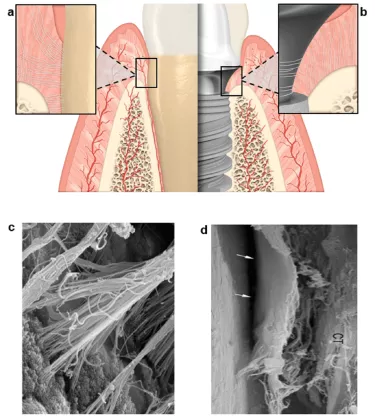
Connective tissue on natural teeth anchor to the cementum using perpendicular collagen fibers called Sharpey’s fibers (Figure 1a, c).2,3 These fibers ensure a tight seal between the soft tissue and tooth, and contribute mechanically to strengthen the barrier from bacterial infiltration or other foreign body penetration. Conversely, collagen fibers adhere parallel or circumferentially on the transgingival region of abutment surfaces (Figure 1b, d), resulting in a weaker connective interface.2-4 The modest adhesion of peri-implant fibers cannot withstand mechanical forces as efficiently as the strong attachment of Sharpey’s fibers of natural teeth.1,5
Moreover, in natural teeth, the blood supply to periodontal tissue is provided by the periodontal ligament, periosteum, and connective tissue. Dental implants do not benefit from the periodontal ligament vasculature and, therefore, the surrounding tissue has a limited blood supply compared to natural teeth.1,5 Additionally, the surgical trauma of implant placement causes fibrotic tissue to form at the implant-abutment complex, which further limits vascularization to adjacent soft tissue.1,6 This reduced vascularization can affect the defense of the peri-implant soft tissue against bacterial infiltration.1
To aid in the prevention of peri-implant disease, this fragile peri-implant soft tissue barrier must be carefully maintained through good oral hygiene and proper soft tissue handling using soft-tissue-friendly prosthodontics.
While there are many factors that contribute to the success or failure of implant-supported restorations, the quality and the quantity of existing or augmented peri-implant mucosa has a critical role to play. In addition to the features discussed above, scientific consensus supports soft tissue site development (tissue grafting) to improve implant outcomes. Especially when the initial soft tissue vertical thickness is less than 2 mm and when the keratinized oral epithelia is limited.7,8
Prosthodontic considerations for soft tissue management
When using a two-stage procedure with bone-level implants, disconnecting the healing abutment can rupture the adherent epithelial and connective tissue from underlying tissue (Figure 2). The resulting wound exposes the connective tissue (Figure 2a). Repeated abutment disconnection on bone-level implants could harm the mucosal barrier and lead to an apical migration of the biological width and consequently early peri-implant bone loss.9,10 Abrahamsson et al. originally described this observation in a canine study using a platform-matched connection.10 Similar results were seen for implants with platform-shifting and internal connection but to a lesser extent.11,12 Therefore, the number of abutment disconnections should be minimized to prevent damage to the mucosal barrier and peri-implant bone resorption.
Clinical recommendations and the On1 concept as a solution
According to available data, it would be beneficial to minimize disruption of the soft tissue after implant placement. Unfortunately, with conventional bone-level implants, the soft tissue can be disrupted at numerous points during treatment, such as healing cap removal, impression coping, and definitive prosthesis delivery (Figure 3a). For this reason, the soft-tissue-friendly final abutment at time of implant placement approach was developed. With this method, the final abutment is installed at implant placement and left undisturbed throughout the treatment process.9,13,14,15
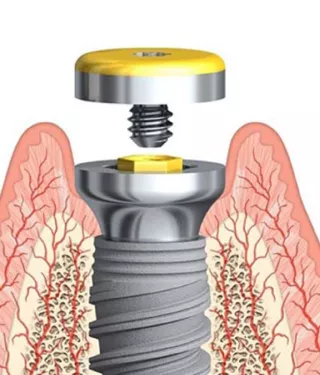
Based on these principles of peri-implant soft tissue preservation, Nobel Biocare developed the On1 concept. The On1 concept has a two-piece abutment with a multi-compatible base that is fixed at implant placement (Figure 3b).
The base design incorporates platform shifting and a conical connection to enhance soft tissue organization and to maintain torque throughout abutment exchanges. To provide maximum flexibility the On1 concept is compatible with several bone-level implants. The base has a transgingival design that is available in two heights to accommodate different soft tissue thicknesses. The system includes healing caps and impression copings, temporary and final abutments, which offers full flexibility in terms of prosthetic procedures. These features incorporate the benefits of both tissue- and bone-level implants into one system.
Conclusion
Peri-implant soft tissue maintenance is important for long-term health of tissues surrounding implants and restorations. Both clinicians and manufacturers should be conscious of the anatomical and prosthodontic factors that affect the peri-implant soft tissue interface and ultimately the success of the implant. Eliminating the need to disrupt the peri-implant soft tissue interface through multiple disconnections of the abutment from the bone-level implant could also minimize undue risk to implant health.
The use of a two-piece abutment with a fixed base, such as that of the On1 base, when coupled with a bone-level conical connection implant and platform shifting, addresses many of these considerations compared to conventional one-piece abutments. This concept promotes an undisturbed peri-implant soft tissue interface while retaining prosthodontic flexibility and ease of use.
More to explore
Dr. Lambert is a Head of Clinic, Department of Periodontology and Dental Surgery and Associate Professor, Dental Biomaterial Research Unit, University of Liège and Former President of the Belgian Society of Periodontology.
Some products may not be regulatory cleared/released for sale in all markets. Please contact the local Nobel Biocare sales office for current product assortment and availability.
References
1. Wang, Y., Zhang, Y. & Miron, R. J. Health, Maintenance, and Recovery of Soft Tissues around Implants. Clin Implant Dent Relat Res 18, 618-634, (2016).
2. Berglundh, T. et al. The soft tissue barrier at implants and teeth. Clin Oral Implants Res 2, 81-90, (1991).
3. Schupbach, P. & Glauser, R. The defense architecture of the human periimplant mucosa: a histological study. J Prosthet Dent 97, S15-25, (2007).
4. Tomasi, C. et al. Morphogenesis of peri-implant mucosa revisited: an experimental study in humans. Clin Oral Implants Res 25, 997-1003, (2014).
5. Atsuta, I. et al. Soft tissue sealing around dental implants based on histological interpretation. J Prosthodont Res 60, 3-11, (2016).
6. Moon, I. S., Berglundh, T., Abrahamsson, I., Linder, E. & Lindhe, J. The barrier between the keratinized mucosa and the dental implant. An experimental study in the dog. J Clin Periodontol 26, 658-663, (1999)
7. Akcali, A. et al. What is the effect of soft tissue thickness on crestal bone loss around dental implants? A systematic review. Clin Oral Implants Res (2016).
8. Puisys, A. & Linkevicius, T. The influence of mucosal tissue thickening on crestal bone stability around bone-level implants. A prospective controlled clinical trial. Clin Oral Implants Res 26, 123-129, (2015).
9. Degidi, M., Nardi, D. & Piattelli, A. One abutment at one time: non-removal of an immediate abutment and its effect on bone healing around subcrestal tapered implants. Clin Oral Implants Res 22, 1303-1307, (2011).
10. Abrahamsson, I., Berglundh, T. & Lindhe, J. The mucosal barrier following abutment dis/reconnection. An experimental study in dogs. J Clin Periodontol 24, 568-572, (1997).
11. Rodriguez, X. et al. The effect of abutment dis/reconnections on peri-implant bone resorption: a radiologic study of platform-switched and non-platform-switched implants placed in animals. Clin Oral Implants Res 24, 305-311, (2013).
12. Becker, K., Mihatovic, I., Golubovic, V. & Schwarz, F. Impact of abutment material and dis-/re-connection on soft and hard tissue changes at implants with platform-switching. J Clin Periodontol 39, 774-780, (2012).
13. Canullo, L., Bignozzi, I., Cocchetto, R., Cristalli, M. P. & Iannello, G. Immediate positioning of a definitive abutment versus repeated abutment replacements in post-extractive implants: 3-year follow-up of a randomised multicentre clinical trial. Eur J Oral Implantol 3, 285-296, (2010).
14. Koutouzis, T., Koutouzis, G., Gadalla, H. & Neiva, R. The effect of healing abutment reconnection and disconnection on soft and hard peri-implant tissues: a short-term randomized controlled clinical trial. Int J Oral Maxillofac Implants 28, 807-814, (2013).
15. Molina, A., Sanz-Sanchez, I., Martin, C., Blanco, J. & Sanz, M. The effect of one-time abutment placement on interproximal bone levels and peri-implant soft tissues: a prospective randomized clinical trial. Clin Oral Implants Res, (2016).


