Anterior implant esthetics
Dr. Bernard Touati provides insight into anterior soft tissue management techniques and procedures.
Past President of the European Academy of Esthetic Dentistry, Dr. Bernard Touati is also a member of the American Academies of Restorative Dentistry and Esthetic Dentistry. He practices in Paris, France, and lectures around the world on practical and innovative dental procedures. The following article provides a condensed version of a lecture he held at the Nobel Biocare Global Symposium in New York City last June.
Dr. Bernard TouatiIn the following article, I will do my best to explain the main factors influencing hard and soft tissue remodeling around implants and make suggestions on how to achieve optimal integration in the esthetic zone.
Among other aspects of treatment, I will cover diagnostics, treatment planning and risk assessment; ideal 3D bone-level implant placement, the relevance of good hard tissue volume and architecture as well as the importance of thick and stable soft tissue in the trans-mucosal zone.
When we deal with dental implants in the anterior region, we are looking for more than osseointegration. We—doctors and patients alike—are looking for optimal soft tissue integration. We are looking for the perfect pink score. In the anterior region, esthetic perfection is not a choice but an obligation. Patients want to have their peri-implant soft tissue mimicking the soft tissue around natural teeth.
There are, of course, many differences between teeth and implants. When we produce restorations based on natural teeth, the gingiva is only dealing with the margin of the crown. We locate our margin at the gingival level or intra-sulcular, but not transmucosally. When we deal with implants, on the other hand, we need to take into consideration the mucosal barrier, and the mucosal barrier is quite different on implants than on teeth for a variety of reasons.
The problem is that when we want to do something transmucosally—for the abutment or at the neck of the implant—we need to have the soft tissue adhering to the prosthetic surface of the implant. This is different than working with natural teeth because it involves many biological factors. To achieve harmonious soft tissue integration, we obviously have to take into account all the biological, functional and esthetic factors.
And not just in two dimensions!
We have to remember that our work will not be evaluated by the 2D photographic images we use to document the treatment, but in the homes, on the streets and at the workplaces where our patients live their day-to-day lives. We thus need to achieve 3D integration. We need to have the scalloping, the volume, the papillae, the texture, the color and the absence of scars that are characteristic of healthy, natural teeth.
Five major factors
The main factors that influence tissue remodeling around implants can be organized into five categories: anatomically-, surgically-, implant-, patient- and prosthetics-related.
Among the anatomical considerations are the tissue biotype, the thickness of the bone plates, the thickness of the soft tissue and the lack of attached gingiva. I can testify from experience that the tissue biotype and the thickness of the tissue are really decisive to optimal outcomes.
Surgical factors include the implant position in three dimensions, the choice of the flap or flapless approach, and the kind of soft tissue augmentation that has been carried out. Other factors include bone desiccation, countersinking, bone compression and—not least of all—the extraction technique used.
Implant design is also important, of course. The design of the neck, the surface properties of the implant and the type of connection can all be decisive. Questions that become interesting in this context include, “Do we have platform shifting available?” or, “Can the implant be maneuvered during insertion, when necessary, in order to ensure optimal placement?”
We also need to remember that every patient has a specific set of characteristics that influence remodeling. Do they smoke? Do they have good healing potential? Immune factors need to be considered, as well as the patient’s willingness and ability to maintain good oral hygiene.
There are also a great number of prosthetic factors that impact remodeling. The final abutment design, the biomaterial from which they are made, the abutments’ surface properties, connection and fit are all important factors that contribute to success. Abutment connections—and disconnections—need to be taken into consideration as well as choices concerning immediate provisionalization and the submergence profile, emergence profile and the restoration anatomy. We need to be careful about deleterious excess cement (if we have not chosen screw retention, of course) and must take into account good occlusion to prevent excessive load.
Given all these factors—and I have only listed the main ones in the table at the bottom—I have constructed a roadmap for optimal integration in the esthetic zone.
I’ll guide you through the first half of my roadmap here on the pages of Nobel Biocare News, and if you would like to find out where the final steps can lead you, you’ll find a link with a QR code at the end of this article that will take you to the full roadmap video online.
Diagnose, plan and assess
To plan for a successful anterior solution, we need to assess risk factors via 3D visual inspection, probing and employing radiographs. Visually, we can see deformities such as concavities. Probing, we can see where the bone is and we can also probe at the level of the adjacent teeth to assess the periodontium.
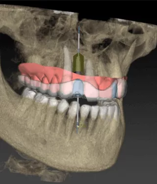
Cone Beam Computed Tomography (CBCT) is an invaluable 3D tool. When connected to the NobelClinician Software, it provides us with an enormous amount of information useful in the decision-making process. It shows us, for example, whether we have a thin buccal plate or a thick one. And this makes a very big difference. Also the volume and the architecture of the site become clear when viewed with the NobelClinician Software.
Assessing the thickness of the soft tissue is also possible via (CB)CT.When the patient wears radiographically transparent lip retractors during the imaging process, (which keeps the lips apart from the teeth and retracts the tongue), the resulting (CB)CT image renders the soft tissue in light gray. Then if we want to harvest some soft tissue from the palate, for example, this technique allows us to objectively measure the tissue available.
This technique also lets us know the patient’s biotype, thin or thick. A delicate biotype will ordinarily require connective tissue grafting, but a thick one generally indicates stable tissue that is forgiving of minor mistakes. When facing thin and moderate soft tissue situations, we will need to be more invasive, go through procedures for soft tissue enhancement, grafting procedures, etc.
(Editor’s note: See the cover picture for a glimpse of NobelClinician’s new smart fusion technology merging (CB)CT scans and tissue information from the NobelProcera 2G Scanner.)
Ideal implant placement
The first thing we have to think about is where we are going to place the implant. The ideal 3D position is very critical because even a little deviation can impact the esthetic outcome.
Using NobelActive, I can make the small adjustments during insertion that ensure optimal 3D placement (which is essential in anterior cases). At the same time, this implant provides excellent initial stability—and in anterior cases we need to reach both initial stability and ideal three-dimensional positioning.
The real problem is the transversal. We want to insert our implant more towards the palatal, because if we leave too much inclination, we run the risk of reducing the thickness of the buccal plate, which in almost all cases is already very thin.
The more the implant allows you to play with the position of the implant—in order to put it in solid bone—the better suited the implant is to situations like these. With a little extra room between the buccal plate and the implant, you will have space to fill in later with bone augmentation material.
Ensuring ample hard tissue volume and good architecture
At this point, we are dealing with where the bone is, and how to make the most of it. Again, we really do need to keep in mind how thin the buccal plate ordinarily is.
With teeth, we have Sharpey’s fibers, we have the blood supply of the periodontium, we have stimulation, and even though we don’t have much, if any, cortical on the buccal, the soft tissue still stays in place. With an implant, on the other hand, we run the risk of fenestration through this thin bone if we position implants in the same orientation as natural incisor roots.
The buccal socket wall is predominantly composed of bundle bone. The lack of stimulation and function in the absence of Sharpey’s fibers may explain the remodeling of this wall while the lingual one has more lamellar bone.
The buccal plate often collapses quickly when we extract a tooth—partly because it is thin, and partly because it is mostly composed of bundle bone. Because an implant does not have a periodontium and therefore lacks vascularisation, we have a ready explanation as to why we have more remodeling on the buccal side as opposed to the lingual side.
In 60 percent of anterior cases, buccal bone plates are less than 0.5 mm thick (and we really need 2 mm to get the job done). If we remember these values, we will understand the entire strategy of slightly angulating the implant in the anterior aspect.
When building a multiple-unit anterior restoration on natural teeth, we still have soft tissue, and the soft tissue is quite stable. But once anterior teeth are extracted, we will almost certainly have to reorient “the root,” inserting the implant palatally.
The good news is that with the (CB)CT—especially when used in conjunction with NobelClinician Software—we can objectively assess that we are in the right position first, making sure that a gap exists between the implant and the buccal plate. This way we can take steps to thicken the buccal bone plate zone and thus provide a safe situation for the future.
The key factors for good esthetic results at anterior extraction sites are the integrity of the buccal plate and the thickness of the soft tissue.
If those two parameters are promising, we are going to find ourselves on the safe side, and are likely to succeed. Of course, in terms of the 3D architecture of the soft tissue and recapturing interdental papillae, the health of the periodontium of the adjoining teeth is important, and probing gives us solid information.
Bone grafting
We can use bone augmentation materials in the jumping gap (i.e. the osteogenic “jumping distance,” which is the gap between the implant body and the alveolar wall). In cases where we have a big defect, we can carry out guided bone regeneration (Editor’s note: please see pages 10–11 in this issue). Adding connective tissue on top of this, which we often do, brings greater thickness to the tissue, which provides greater mechanical resistance and leads to increased blood supply.
In cases where there is no buccal bone post-extraction, we firstly need to recreate a complete socket, which provides a favorable 3D situation for the insertion of an implant and is well within widely-practiced, well-accepted reconstructive protocols.
Establishing thick and stable soft tissue
This brings us to the soft tissue. We need to have it thick and stable, especially in the transmucosal zone. Stability makes for good esthetics.
Post-extraction remodeling is inevitable. Today there is no magical way to totally counteract the post-extraction remodeling—it’s biological—but we can compensate for it. One way to compensate is to thicken the soft tissue and also to regenerate the bone, when necessary.
A connective tissue pouch can be used, both horizontally and vertically. Mucosal enhancement can be realized through connective tissue graft(s).
Not least of all, we can manipulate the soft tissue via subtle changes in the design of the prosthetics.
First, we want to see some concavity transmucosally at the abutment level (“curvy”) and/or “platform switching” in order to thicken the mucosa, creating a virtual “o-ring” of soft-tissue. On the other hand, proximally the prothetic restorations need to display some convexity in order to gently push the tissue and to keep the interdental papillae.
The vertical position of the implant in relation to the mucosa is very important. This makes it possible for us to play with the emergence profile and then shape the marginal mucosa with the emergence bulk. Adding composite material incrementally, we can very successfully guide the marginal mucosa. This careful step-by-step process takes a substantial amount of time, but gives beautiful results.
This covers a little more than half of my roadmap for optimal integration in the esthetic zone. The effects of early or immediate loading, experience of the perfect provisional for soft tissue conditioning, and how best to use zirconia-based concave abutments and screw-retained crowns are all explored online in Part Two. See “More to explore” below.
Major factors affecting hard and soft tissue remodeling around implants.
Anatomically-related
– Tissue biotype
– Thickness of bone plates
– Thickness of soft tissue
– Lack of attached gingiva
Surgically-related
– Implant 3D position
– Flap elevation or flapless surgery
– Soft tissue augmentation
– Bone desiccation
– Countersinking
– Bone compression
– Extraction technique
Implant-related
– Design (macro, micro)
– Surface properties
– Type of connection
– Built-in platform shifting or equivalent
Patient-related
– Hygiene
– Maintenance
– Tobacco
– Healing
Prosthetics-related
– Final abutment design
– Type of abutment connections
– Provisional abutment: biomaterial, design
– Immediate provisionalization
– Submergence profile
– Emergence profile
– Restoration anatomy
– Possible excess cement and retention
– Occlusion, excessive load
More to explore
– Full lecture: Bernard Touati: Anterior implant esthetics - optimal soft tissue integration | FOR.org